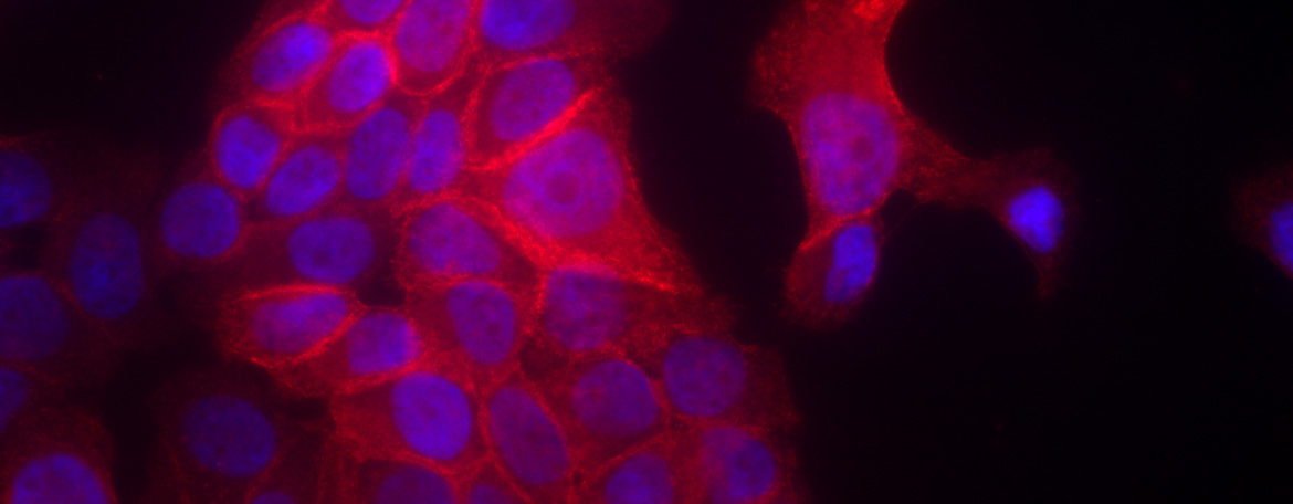
Students interested in understanding the molecular mechanisms that underlie human disease will find a home in the Molecular Biology and Biochemistry (MBB) Graduate Program. From cancer to host-pathogen interactions, our students study the proteins and pathways involved with an eye toward improving disease diagnosis, prevention and treatment.
MBB students are affiliated with the Department of Molecular Biology and Biophysics, which provides a rigorous, yet supportive community of faculty, students, and staff to guide them through the Ph.D. degree process.
The primary goal of the MBB Graduate Program is to train students for the broad range of careers available in biomedical science. Whether the graduate pursues a career in academic research, biomedical industry, teaching, government or any of the other careers now available to Ph.D.s in biomedical science, we have attempted to prepare them with a solid base of knowledge, critical thinking skills and the confidence in their abilities to be successful. Graduates are expected to have demonstrated a high degree of competence in research, as judged by publications in first-rank journals. They will have developed essential skills in identifying important research problems, planning appropriate experimental approaches, and effectively communicating their research results and their significance both orally and in written form. The success of our students in these areas is exemplified by the number of first-author publications in quality scientific journals and awards received both internally at UConn Health and from national and international conferences and societies.
Categories of faculty research:
- Microbiology and Infectious Diseases
- Structural Biology and Biophysics
- Computation and Modeling
- Cellular Pathways
- Cancer Biology
For information on requirements for completion of the Biomedical Science Ph.D. degree in the MBB Area of Concentration, please see the Graduate Program in Molecular Biology and Biochemistry Student Handbook. The handbook lists suggested courses, preliminary exam requirements, thesis exam requirements and other details about the program.
*Admissions Note*
The GRE General Exam is no longer required NOR considered for admission to the Biomedical Science Ph.D. program.
The application for Fall 2024 will open in mid-September. The deadline for application, application fee, and supporting materials for Fall 2024 is December 1, 2023. For detailed application information, please visit our Admissions Process page.